

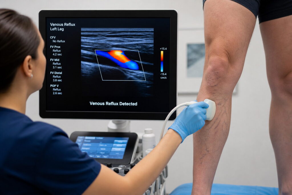
Duplex ultrasound is one of the most important tools in modern vein care. It allows clinicians to see not only the structure of the veins but also how blood is actually flowing through them in real time. This makes it essential for diagnosing venous reflux and creating a precise, effective treatment plan.
What Is Venous Reflux?
Venous reflux occurs when the valves inside your veins stop working properly. Instead of blood flowing upward toward the heart, it flows backward and pools in the legs. Over time, this can contribute to symptoms such as:
- Leg heaviness or fatigue
- Swelling in the ankles or calves
- Visible varicose veins
- Aching or throbbing discomfort
- Skin changes or discoloration in advanced cases
Identifying where reflux is happening is key to treating the underlying problem—not just the visible symptoms.
How Duplex Ultrasound Works
Duplex ultrasound combines two imaging methods:
- B-mode ultrasound: Shows the structure of the veins
- Doppler ultrasound: Shows the direction and speed of blood flow
Together, they allow your provider to see both anatomy and function at the same time.
During the exam, a small handheld probe is moved along the skin while images appear on a monitor. The Doppler function highlights blood flow in color, making it easy to detect abnormal backward flow.
How Venous Reflux Is Identified
One of the key advantages of duplex ultrasound is its ability to actively test valve function. Providers may apply gentle pressure or have you change position to observe how blood moves through the veins.
Reflux is identified when:
- Blood flows in the wrong direction
- Flow continues backward after pressure is released
- Vein valves fail to close properly
This real-time feedback makes duplex ultrasound the most accurate non-invasive method for diagnosing venous insufficiency.
How Ultrasound Guides Treatment Planning
Duplex ultrasound is not just a diagnostic tool—it directly shapes your treatment plan. Once reflux is mapped, your provider can:
- Identify exactly which veins are affected
- Determine the severity of valve dysfunction
- Choose the most appropriate treatment method
- Target only the problematic veins
This precision ensures treatments are more effective and less invasive, avoiding unnecessary procedures on healthy veins.
Why It’s Considered the Gold Standard
Duplex ultrasound is widely considered the gold standard for vein evaluation because it is:
- Non-invasive and painless
- Free of radiation
- Highly accurate in detecting reflux
- Able to assess both structure and blood flow
- Essential for insurance approval of many vein procedures
It provides the foundation for safe, personalized vein care.
From Diagnosis to Results
Once venous reflux is clearly mapped, treatment can be tailored to your specific condition. Because the ultrasound identifies the exact source of the problem, procedures can be performed with precision—leading to better outcomes and faster symptom relief.
Final Takeaway
Duplex ultrasound plays a critical role in modern vein treatment. By revealing both vein structure and blood flow, it allows clinicians to accurately detect venous reflux and design highly targeted treatment plans.
If you are experiencing symptoms such as leg pain, swelling, or visible veins, a duplex ultrasound is the first step toward understanding the cause—and finding the right solution.